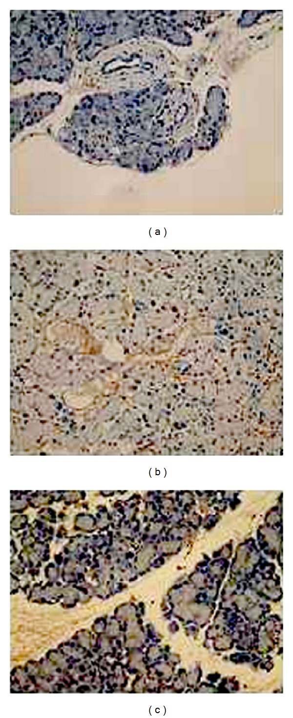
Figure 3.

ICAM-1 immunohistochemistry in pancreases samples. (a) Control group, (b) model group, and (c) treatment group. In weakly staining, positively stained cells showed a brown-yellow pigment in control group. The model group exhibited the strong positive expression of ICAM-1 with lots of brown-yellow stained particles in cells, while in treatment group the change was not remarkable.
