Figure 1.

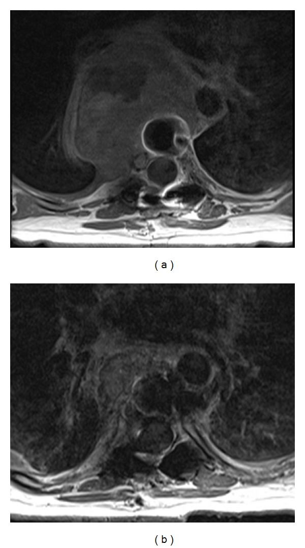
Axial T1 weighted postcontrast computed tomographic images show a large mid-thoracic spine mass (a) before and (b) 10 months after denosumab therapy.

Axial T1 weighted postcontrast computed tomographic images show a large mid-thoracic spine mass (a) before and (b) 10 months after denosumab therapy.