
Figure 3.
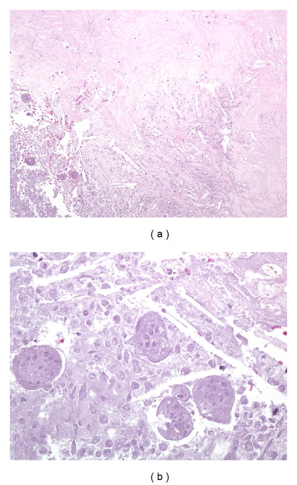
Representative photomicrographs of the paraspinal mass after denosumab therapy. At low magnification ((a); 100x, hematoxylin and eosin), extensive necrosis is apparent with abundant cholesterol clefts (center) and surrounding fibrosis (top). Residual tumor is focally recognizable but is entirely necrotic (lower left), and no viable tumor is present. At high power ((b); 400x, hematoxylin and eosin), “ghosts” of necrotic multinucleated osteoclastic giant cells are present among necrotic mononuclear tumor cells.