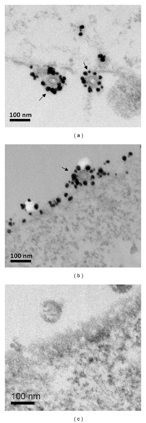
Figure 2.

Cell surface immunogold labeling and electron microscopy of infected cells. Electron microscopy of thin sections of cells co-infected with (a) rBVs expressing tM2e and M1; (b) rBVs expressing tFliC, tM2e, and M1; or (c) rBVs expressing M1 only. Surface immunolabeling was done prior to fixation, embedding, and sectioning of the infected cells at 48 h postinfection. The primary antibody was a mouse anti-M2e antibody at 1 : 100 dilution, and the secondary antibody was 10 nm gold-conjugated goat anti-mouse antibody at 1 : 100 dilution. The particles show a prominent layer of surface spikes (bar = 100 nm).
