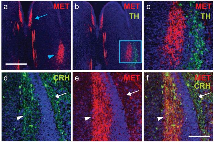
Fig. 5.
MET is expressed by neurons in Barrington’s nucleus. Immunostaining for MET, tyrosine hydroxylase (TH), and corticotropin releasing hormone (CRH) at E16. (a-c) Arrowhead denotes MET+ neurons situated medial to the TH+ locus coeruleus. Note the absence of double-labeled neurons. The arrow in (A) indicates MET+ neurons in the dorsal raphe. (d-f) In contrast, immunostaining for CRH and MET revealed co-localization in neurons residing within Barrington’s nucleus (arrowhead), but not in the more laterally situated locus coeruleus (arrow). Scale Bar: top = 30 μm; bottom = 10 μm.

