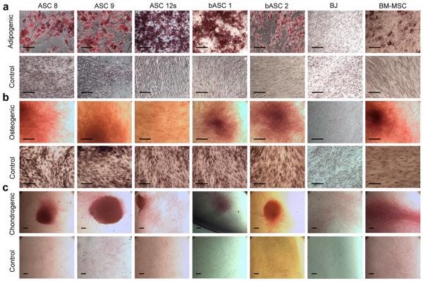
Figure 2. ASCs differentiate into fat, bone, and cartilage.
ASC 8, ASC 9, ASC 12s, bASC 1, bASC 2, BJ, and BM-MSCs were tested for their ability to differentiate into 3 mesenchymal lineages. Cells grown in control media (standard growth media) served as negative controls. (a) Adipogenic differentiation was visualized as lipid droplets in a significant proportion of cells (~70%) after 14 days. Cultures were fixed and stained with Oil Red O to specifically highlight lipid formation indicative of adipocytes. (b) All 5 cell strains were incubated at confluency in either osteogenic media or control media for 2–3 weeks to induce osteogenic differentiation. Once cells visually formed aggregates reminiscent of bone nodules and produced significant amounts of calcium deposits (2–3 weeks), cultures were fixed and stained with Alizarin Red S to detect calcium production, which is representative of osteoblast differentiation. (c) Following micromass pellet formation (see Experimental Procedures), all cells were continuously cultured in either chondrogenic induction media or control media for 30 days. The entire pellet was processed and stained with Safarin O to permit detection of GAG protein production indicative of chondrogenic differentiation. All images in (a) and (b) were captured at 10x magnification, while images for (c) were captured at 4x magnification. The scale bar in each indicates 100μc.

