
Figure 4.
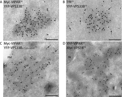
Ultrastructual localizations of VPS33B, VPS33B(c.1225G>C), VIPAR, and VPS18 constructs. Transmission electron micrographs of ultrathin cryosections of HEK293 cells. A, B: Cells cotransfected with YFP-tagged VPS33B and Myc-tagged VIPAR were immunogold stained with anti-GFP/YFP (15 nanometer gold) and anti-Myc (10 nanometer gold) (A), or anti-GFP/YFP (15 nanometer gold) and anti-TfR (10 nanometer gold) (B). Colocalization was observed on endosome (E)-associated tubular–vesicular membranes typical of recycling endosomes, which was confirmed by the presence of TfR (arrows in B). (C) Cells co-overexpressing YFP-VPS33B(c.1225G>C) and Myc-VIPAR were immunogold labeled for anti-GFP/YFP (15 nanometer gold) and Myc (10 nanometer gold). The two proteins colocalized in cytosolic aggregates with partial staining of VPS33B(c.1225G>C) on nearby vesicles (arrows). D: Cells co-overexpressing YFP-tagged VIPAR (labeled with anti-GFP; 10 nanometer gold) and Myc-tagged VPS18 (labeled with anti-Myc, 15 nanometer gold) showed colocalization in cytosolic aggregates. E, endosome; N, nucleus; PM, plasma membrane. Scale bars, 200 nm.