Figure 8.

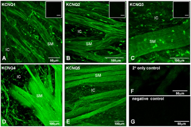
Detection of KCNQ gene expression by immunohistochemistry. (A–E) Whole-mount preparations of guinea pig detrusor were incubated with antibodies to KCNQ1-5 subtypes. Immunoreactivity was detected in both smooth muscle bundles (SM) and in IC adjacent to and between the smooth muscle bundles for the five KCNQ subtypes tested. Inset micrographs in (A–C) show minimal immunofluorescence in pre-absorption controls for KCNQ1-3; the scale bars indicate 50 μm. (F) Micrograph of secondary antibody only control slide, showing minimal immunofluorescence. (G) Micrograph of negative control (no antibodies) demonstrating absence of autofluorescence.
