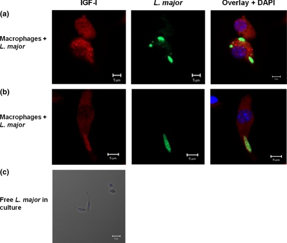
Figure 1.

Detection of IGF-I within RAW 264·7 macrophages following infection with Leishmania major promastigotes. Co-localization of IGF-I and Leishmania was measured using immunofluorescence. Anti-IGF-I antibody (recognized by the secondary antibody AlexaFluor-546, shown in red) and anti-Leishmania antibody (recognized by the antibody AlexaFluor-488, shown in green) were used to label cells (a, b). Free L. major promastigote culture immunofluorescence was measured using anti-IGF-I and the secondary antibody AlexaFluor-546 (c). No IGF-I staining was observed (red). 4′,6-diamidino-2-phenylindole (DAPI, shown in blue) was used to stain nuclei. Images were captured with a Leica LSM510 confocal microscope with a 63× objective and oil immersion.
