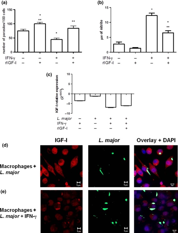
Figure 2.

Parasitism, nitric oxide (NO) production, IGF-I mRNA and IGF-I protein expression in RAW 264·7 cells that were infected with Leishmania major promastigotes. (a) Parasitism (shown as the number of parasites per 100 cells), (b) NO as nitrite levels evaluated in culture supernatants and (c) IGF-I mRNA expression (calculated as the ratio in relation to the control group without stimulation,  ) with and without IFN-γ (200 U/mL) stimulus and with recombinant IGF-I (rIGF-I, 50 ng/mL) stimulus during a 48 h incubation. The data shown are representative of three independent assays. (d, e) Detection of IGF-I by confocal microscopy, using anti-IGF-I antibody (recognized by the secondary antibody AlexaFluor-546, shown in red) and anti-Leishmania antibody (recognized by the secondary antibody AlexaFluor-488, shown in green). 4′,6-diamidino-2-phenylindole (DAPI, shown in blue) was used to stain the nuclei. Images were captured using a confocal Leica LSM510 confocal microscope with a 63× objective and oil immersion. *P < 0·05 (anova and Tukey's test) in relation to the control group. **P < 0·05 (anova and Tukey's test) in relation to the IFN-γ group.
