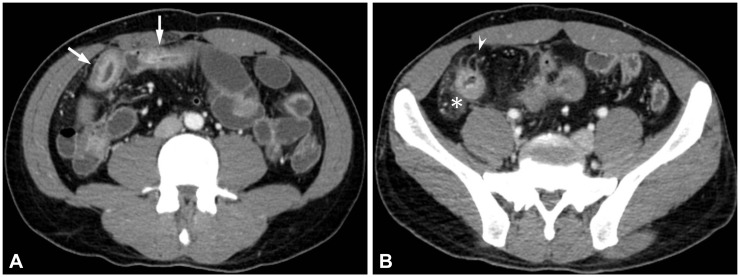
Fig. 1.
Active inflammatory Crohn disease in a 28-year-old man. (A) Axial computed tomography enterography images demonstrate multifocal segmental mural hyperenhancement and trilaminar mural stratification in the ileum (arrows), suggesting active disease. (B) Also note engorged vasa recta (arrowheads) and increased attenuation of the mesenteric fat (asterisk).

