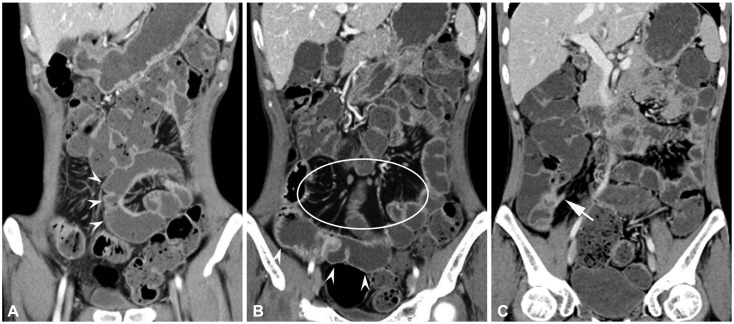
Fig. 3.
Crohn disease in a 31-year-old man. (A, B) Coronal computed tomography enterography images show pseudosacculation along the antimesenteric border of small bowel (arrowheads). (B) Fibrofatty proliferation is also demonstrated in the mesentery (circle). (C) Sinus tract arising in the terminal ileum (arrow) which shows mural hyperenhancement and stratification, suggesting active inflammatory disease, is also noted.

