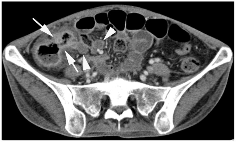
Fig. 8.
Intestinal tuberculosis in a 27-year-old woman. Axial computed tomography enterography image shows enhanced wall thickening involving cecum and terminal ileum with patulous ileocecal valve (arrows). Associated central low attenuated lymph nodes are seen at the ileocecal mesentery, suggesting caseous necrosis (arrowheads).

