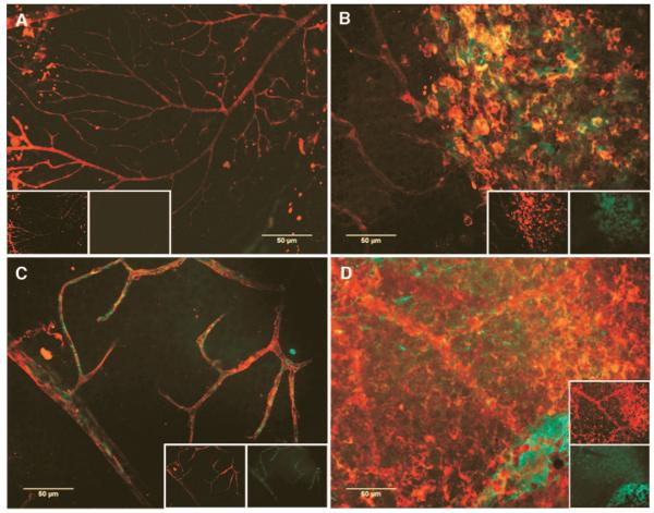
FIG. 3.
Normal huEPC, but not diabetic huEPC, participate in ocular vascular reendothelialization in STZ-diabetic mice. All images are composite red and green channels (shown in respective insets) of epifluorescence digital captures of representative neural retinas. STZ-diabetic (n = 12) and age- and sex-matched normal control mice (n = 12) were given labeled huCD34+ cells from either diabetic (n = 6 diabetic mice, n = 6 nondiabetic mice) or nondiabetic donors (n = 6 diabetic mice, n = 6 nondiabetic mice), and eyes were harvested 48 h later. Panel A is from a normal mouse that received nondiabetic cells. Lack of vascular injury should preclude incorporation of labeled EPC. Note the lack of labeled cells. Panel B is from a normal mouse that received diabetic huEPC. Note the tendency for these labeled cells to form clumps distinct from the vasculature. By contrast, panel C shows extensive incorporation of labeled CD34+ cells from a normal donor in damaged vasculature of a STZ-diabetic mouse. Panel D demonstrates that huEPC incorporation is not solely a function of vascular damage in the recipient, since labeled diabetic CD34+ cells do not integrate into vasculature of a STZ-diabetic mouse, but rather form sheets and clumps separate from the vascular plexus.

