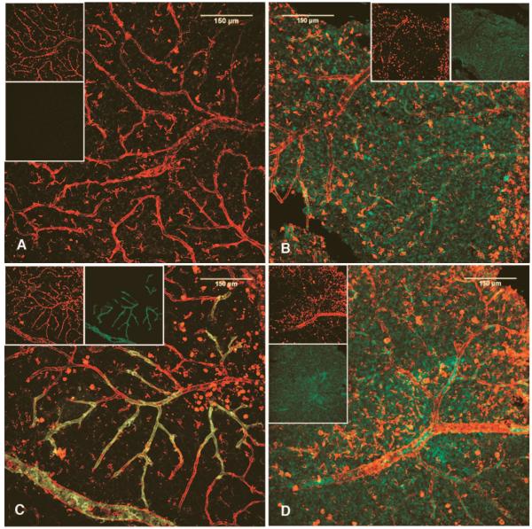
FIG. 4.
huCD34+ EPC of nondiabetic, but not diabetic, origin integrate into damaged vasculature in BBZ/Wor diabetic rats. Labeled cells were given by intravitreous injection to immune suppressed rats (n = 6 nondiabetic rats/nondiabetic EPC; n = 6 nondiabetic rats/diabetic EPC; n = 6 diabetic rats/nondiabetic EPC; n = 6 diabetic rats/diabetic EPC). All images are z-series projections of two-color LSCM, with insets showing respective separate red and green channels. Panel A is from a healthy rat receiving nondiabetic huEPC. Note the lack of labeled cells consistent with lack of vascular injury. Panel B shows that diabetic huEPC in a normal rat eye form a discrete layer without integrating into vessels. Panel C demonstrates clear reendothelialization by labeled normal huEPC into presumably damaged vasculature of a diabetic rat. Panel D shows again that diabetic EPC are incapable of integrating into damaged vasculature, instead forming distinct clumps and layers separate from vessels.

