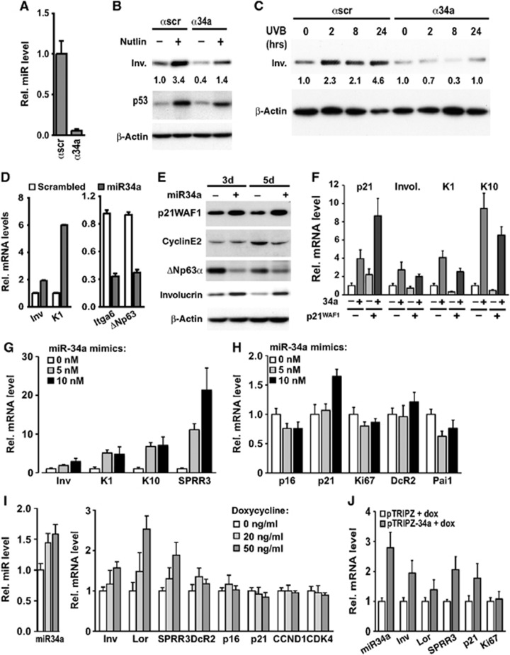
Figure 3.
miR-34a is a positive determinant of keratinocyte differentiation. (A) Proliferating HKCs were transfected for 3 days with miR-34a-specific antagomiRs (α34a) (50 nM) or control scrambled antagomiR (αscr) and levels of mature miR-34a were measured by qRT–PCR using specific Taqman probes. (B) HKCs were transfected as in A and, 24 h later, treated or not with nutlin-3a (10 μM) for additional 48 h. Involucrin and p53 protein levels were analysed by immunoblotting with β-actin as equal loading control. Numbers correspond to folds of induction of involucrin expression relative to untreated control, calculated after densitometric scanning of the autoradiographs and normalization for β-actin expression. (C) HKCs transfected with antagomiR against miR-34a (α34a) or control (αscr) as in A for a total of 3 days were UVB irradiated (50 mJ/cm2) at the indicated times from the end of the experiment, followed by immunoblot analysis for involucrin expression and data quantification by densitometric analysis as in the previous panel. Pattern of p53 protein expression is shown in Supplementary Figure S2J. (D) HKCs transfected for 3 days with miR-34a precursor (+) (25 nM) or scrambled control oligonucleotides (−) were examined for involucrin (inv), keratin 1 (K1), integrin α6 (itga6) and ΔNp63α expression by qRT–PCR with 36β4 for normalization. (E) HKCs at 3 and 5 days after transfection as in the previous panels were analysed by immunoblotting for expression of the indicated proteins. p21WAF1, cyclin E2 and β-actin blots were performed by sequential blotting of the same membrane without stripping, while ΔNp63α and involucrin expression was assessed by a parallel gel/blot with β-actin giving the same pattern of expression. (F) HKCs were stably infected with a lentivirus for doxycycline-inducible expression of p21WAF1/Cip1. Cells were either untreated (−) or treated with doxycycline (500 ng/ml) for 5 days (+) for sustained p21WAF1/Cip1 expression and associated cell cycle arrest. Cells were transfected with miR-34a mimics (25 nM) or scrambled controls for the last 3 days of the experiment. Expression of the indicated genes was assessed by qRT–PCR with 36β4 for normalization. (G, H) HKCs were transfected for 2 days with miR-34a precursor at low concentrations (5 and 10 nM) with scrambled controls used to keep equal total amounts of transfected oligonucleotides. Cells were examined for expression of the indicated differentiation marker (G) and cell cycle and senescence genes (H), by qRT–PCR with 36β4 for normalization. (I) HKCs were stably infected with a lentivirus for doxycycline-inducible expression of miR-34a. Cells were either untreated or treated with doxycycline at the indicated concentrations for 2 days followed by determination of mature miR-34a levels in parallel with expression of the indicated genes by qRT–PCR with 36β4 for normalization. (J) HKCs stably infected with a lentivirus for doxycycline-inducible expression of miR-34a (pTRIPZ-34a) or empty vector control (pTRIPZ) were treated with doxycycline (50 ng/ml) for 4 days, followed by measurement of mature miR-34a levels in parallel with expression of the indicated genes by qRT–PCR as in I.
Source data for this figure is available on the online supplementary information page.

