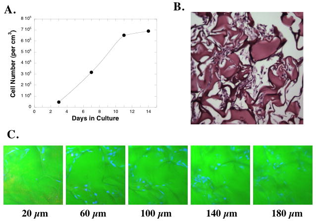
Figure 2. Pulmonary fibroblasts grow within Gelfoam matrices.

(A) NNRLFs were cultured in Gelfoam sponges for the indicated time and cell number was determined after solubilization of the Gelfoam. (B) Histological specimen on NNRLFs in Gelfoam after 14 days in culture. (C) Confocal microscopic images of NNRLFs within Gelfoam sponges. NNRLF-Gelfoam constructs were fixed and cell nuclei stained with TO-PRO (blue) and a z-stack was collected at 20 μm intervals from the top to the bottom of the construct (total width of construct ~3 mm). Samples are shown for the indicated depth.
