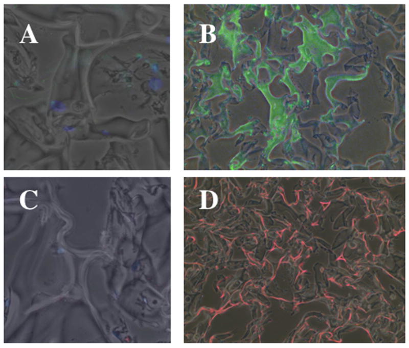
Figure 3. Pulmonary fibroblasts deposit elastin and collagen within Gelfoam matrices.

NNRLFs were cultured in Gelfoam sponges for 14 days, fixed, sectioned and incubated without (A and C) and with primary antibodies to elastin (B), and type I collagen (D), followed by incubation with FITC-linked (A and B, green) or Cy3-linked (C and D, red) secondary antibodies. Secondary antibody alone control samples were visualized using a 20x objective in order to be able to observe the low background fluorescence. The primary antibody samples were observed with a 10x objective so that the distribution of elastin and collagen deposition could be visualized throughout a larger region of the cell-Gelfoam constructs.
