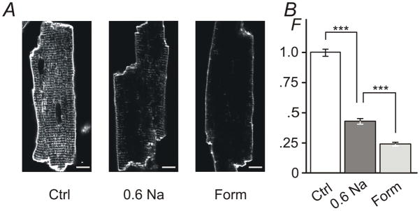
Figure 4. The effect of hypo- and hyper-osmotic detubulation observed with membrane specific dye di-8-ANEPPS.

A, Ventricular myocytes were exposed to 0.6 Na hypo-osmotic or 1.5 M formamide hyper-osmotic solutions for 7 min and 15 min, respectively, returned to normal Tyr solution, labeled with di-8-ANEPPS dye along with control cells (Ctrl; not subjected to osmotic stress) and imaged using confocal microscopy. Ctrl myocytes display strong intracellular fluorescence originating from t-tubular membranes. In contrast, both types of osmotic shock lead to significant reduction of ‘intracellular’ labeling consistent with detubulation. Bar: 10 μm. B, Quantification of ‘intracellular’ fluorescence (not including the outer sarcolemma or intercalated disks; see Methods). The amount of t-tubular fluorescence is reduced to ~43% and to ~24% of that in control myocytes after treatment with 0.6 Na and 1.5 M formamide solutions, respectively. F -Relative fluorescence. Mean fluorescence for Ctrl was set to 1.
