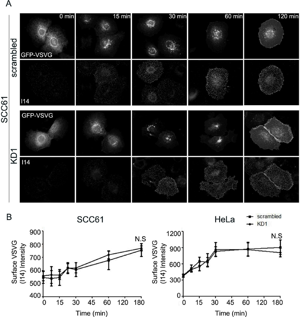
Figure 3. Cortactin-KD does not affect transport of VSV-G to the plasma membrane.
Transport of ts045-VSV-G-GFP (VSV-G) to the plasma membrane was assessed over a 3-h period following a shift of the cells to the permissive temperature of 32°C in SCC61 or HeLa cells expressing either control (scrambled) or cortactin-specific shRNA (KD1). A. Representative images showing total expressed VSV-G (green) and cell surface I14-detected VSV-G (red) in SCC61 cells as a function of time. Images for HeLa cells are shown in Figure S5. B. Analysis of the average surface intensity of I14 VSV-G staining at each time point after the shift to 32°C for SCC61 (left) or HeLa (right) cells. n=3; >20 cells per independent experiment at each time point. N.S. = Not significant. Scale bar = 25 µm.

