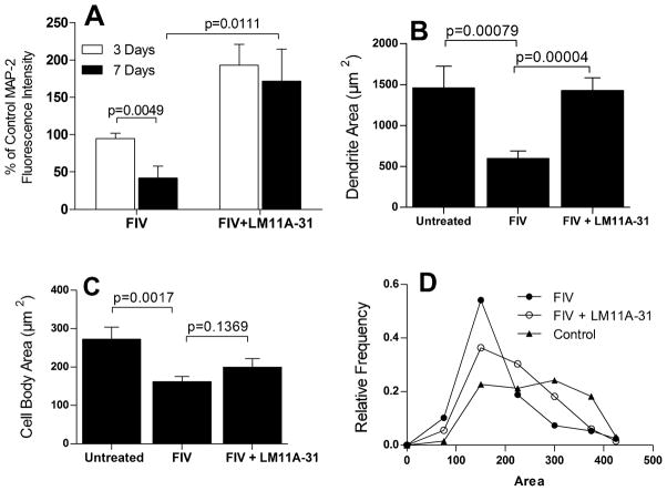
Figure 4.
Quantification of MAP-2 staining relative to untreated controls at 3 and 7 days post-inoculation. A. At 3 days no significant change in average MAP-2 fluorescence intensity from controls was seen whereas by 7 days a 52% decrease was seen (p=0.0049 vs. untreated, n= 6 each). Values are normalized to controls (100%). The FIV-associated decrease in MAP-2 immunoreactivity was completely reversed in the presence of 10 nM LM11A-31 (p=0.0111, FIV vs. FIV+LM11A-31, n=6 each) to a value that was 172% of untreated controls. B. Measurement of the area occupied by MAP-2 stained dendrites at 7 days indicated that the effects of FIV were largely due to a decrease in the dendritic architecture (p=0.00079, n=16) with almost complete prevention of the loss by 10 nM LM11A-31 (p=0.00004, n=11). C. The area of neuronal cell bodies also decreased significantly at 7 days (p=0.0017, n=16) in the FIV-infected cultures but was only partially reversed by LM11A-31 (n=11). D. Characterization of the distribution of neuron cell body areas illustrated that LM11A-31 significantly shifted the distribution toward a broader profile with larger cells (X2 =27.5, df=6, p<0.01, 198–244 neurons), similar to untreated neurons.

