Figure 6.
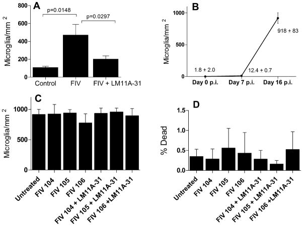
Microglial changes in response to FIV and LM11A-31. A. Neural cultures inoculated with 105 TCID50 FIV showed a significant increase in microglia after 7 days (p=0.0148, FIV vs. Control, n=15 and 11, respectively) which was largely reversed in the presence of 10 nM LM11A-31 (p=0.0297, FIV vs. FIV+LM11A-31, n=15 and 14, respectively). B. Large increases in microglia typically appeared after prolonged exposure to FIV. A significant increase in microglia was seen seven days post inoculation (Day 7 p.i., n=23) but numbers were still very low relative to neurons in the culture (~5% of the neuron density). By day 16 post-inoculation (n=10), microglia were abundant. C. Inoculation of microglia enriched cultures with FIV or treatment with LM11A-31 in the relative absence of neurons failed to affect density (n=4–6 cultures each). D. Inoculation of enriched microglia enriched cultures with FIV or treatment with LM11A-31 - did not produce significant microglial death (0.2–0.6%, n=4–6 cultures each).

