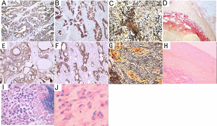
Figure 2.
Examples of histochemical and immunohistochemical stainings of SBO- or xylene-treated sections.
Human tissue samples of colorectal cancer (A, B, E, F), lung cancer (C, G), vermiform appendix (D, H), and hepatitis B virus-infected liver (I, J) were processed with SBO (A-D, I) or xylene (E-H, J). Immunostaining for carcinoembryonic antigen (A, B) and low-molecular-weight cytokeratins (E, F), Gordon Sweet's staining for reticulin fibers (C, G), van Gieson staining for collagen fibers, and Victoria blue staining for hepatitis B surface antigen (I, J) were then performed. Representative staining results are shown. A, B, I, 400 ×; C, 200 ×; D, 100 ×.E,F,J,400 ×;G,200 ×;H,100 ×.

