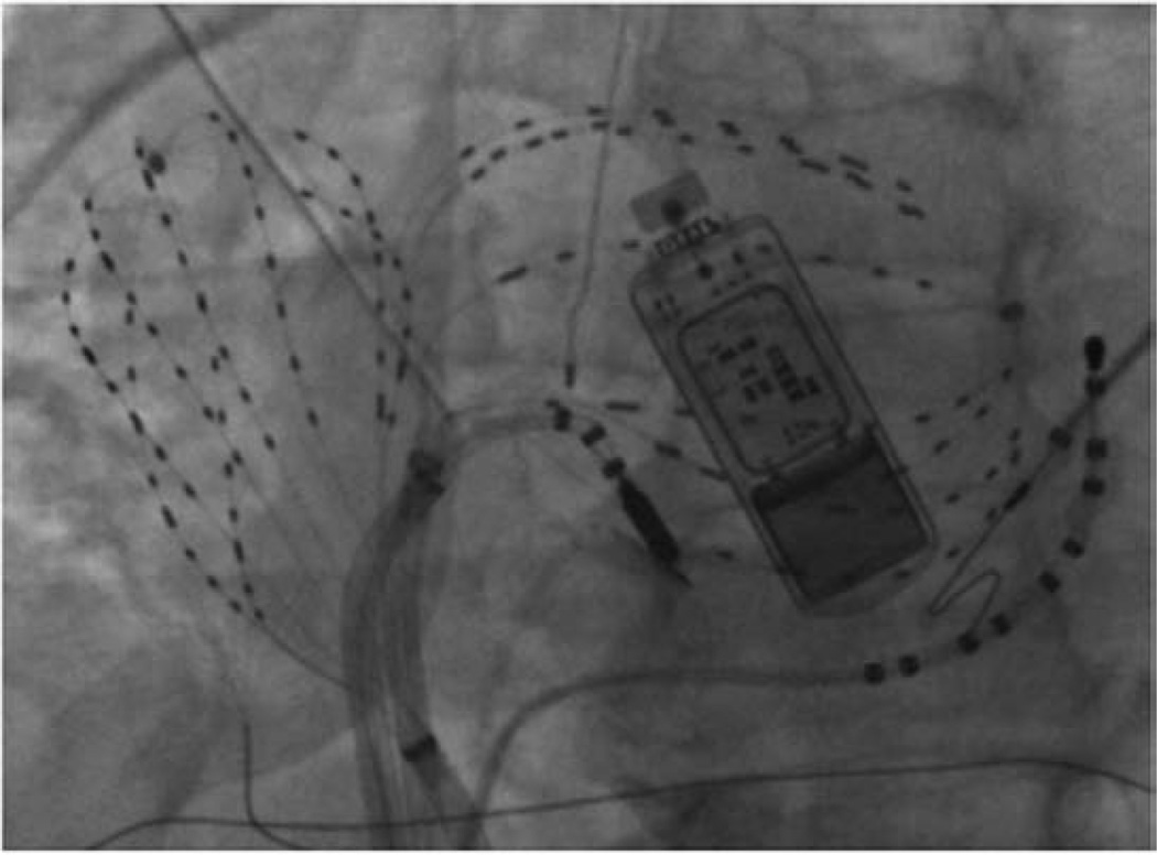
Figure 1.
Contact mapping (focal impulse and rotor modulation mapping) of atrial fibrillation (AF) in both atria. Fluoroscopy shows 64 pole basket catheters used to record AF in the right and left atria, as well as a coronary sinus catheter, an ablation catheter and an esophageal temperature probe. A subcutaneous ECG monitor, implanted preprocedurally to record baseline AF burden, then used postprocedurally to stringently document AF recurrence, is also shown.

