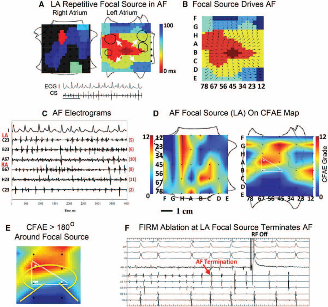
Figure 4.
Stable repetitive atrial fibrillation (AF) focal source, inconsistent relation to complex fractionated atrial electrograms (CFAE). A, Left atrial focal source during AF (time bar = 1 second; Movie III in the online-only Data Supplement); B, activation emanates from focal source to atrium; C, AF electrograms in both atria, referenced to D; D, Panoramic CFAE map showing AF focal source precession (white dots bounded by lines) overlying CFAE (grades 7–10), but also with high CFAE grade remotely; E, CFAE surrounds AF source >180°. F, Focal impulse and rotor modulation (FIRM) ablation at focal source terminated AF toward end of application (RF Off =RF, radiofrequency ablation, OFF), before pulmonary vein isolation. Left and right atrial orientations as in Figure 2A.

