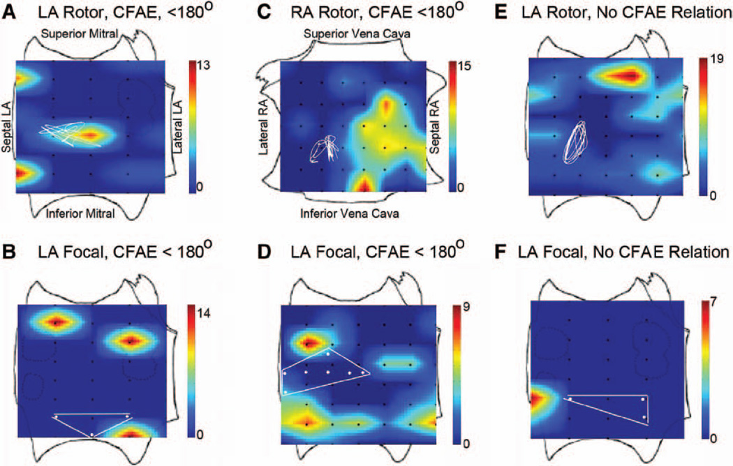
Figure 6.
Human atrial fibrillation (AF) sources showing no overlap and minimal surrounding electrogram fractionation. A, Rotor and (B) focal sources with adjacent but not surrounding (<180°) complex fractionated atrial electrograms (CFAE) in left atria; C, rotor in right atrium and (D) focal source in left atrium with adjacent but not surrounding (<180°) CFAE; E, rotor and (F) focal sources with no clear relation to CFAE. Color scale: CFAE grade, higher numbers (warmer colors) indicate more fractionation. Left and right atrial orientations provided in A and C.

