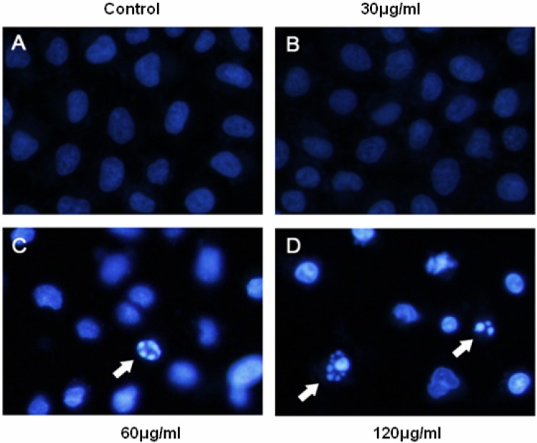
Figure 4.
BEL-7402 cells were incubated with the JMM6 for 24h and stained by Hoechst 33342. The arrows indicate apoptotic cells identified by the Hoechst 33342 staining. (A) the Blank control group. (B) the treated group with 30µg/ml. (C) the treated group with 60µg/ml. (D) the treated group with 120µg/ml.

