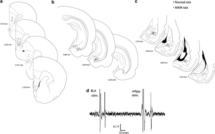
Figure 1.
(a–c) Schematic of coronal sections of the rat brain showing representative placements (circles, for clarity, ∼50%) of recording electrodes in the mPFC (a), and stimulating electrodes in the vHipp (b) and the BLA (c). The brain sections correspond to the atlas of Paxinos and Watson (1986). The numbers correspond to millimeters from bregma. (d) mPFC neuron excited by BLA and vHipp, following stimulation of these regions.

