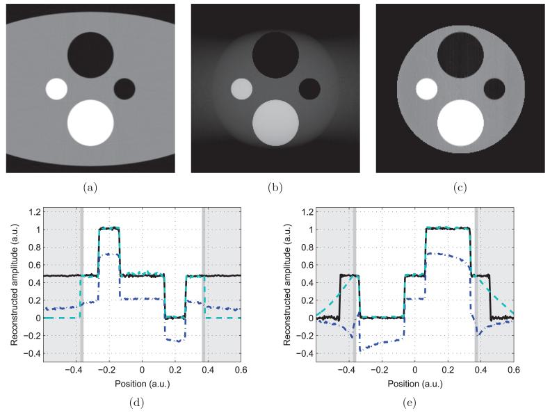
Figure 4.
Reconstructions of the numerical phantom. (a) is the non-truncated FBP reconstruction, (b) is the truncated FBP reconstruction and (c) is the truncated POCS reconstruction. (d) is a vertical profile, while (e) is horizontal. The non-truncated FBP reconstruction is in solid black, the POCS reconstruction is in dashed teal and the truncated FBP reconstruction is in dot–dashed blue. The white background indicates the scanner FOV, the dark gray corresponds to the zone of a priori knowledge—a thin strip at the border of the SFOV—and the light gray area is truncated.

