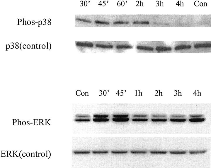
Fig. 1.
Activation of p38 and ERK after exposure to DTDP. Whole-cell extracts of neuronal cultures were harvested at various time points after 10 min exposure to 100 μm DTDP. Proteins were separated on 12% SDS-PAGE gels and probed with antibodies specific to the phosphorylated and nonphosphorylated forms of both p38 and ERK p42/44. Note that there was early increased p38 activation in the first 2 hr after exposure to DTDP and a similar increase in ERK activation within the first 45 min of oxidant exposure. Similar results were obtained in two additional independent experiments.

