Figure 3.
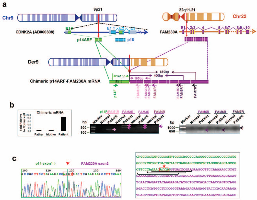
Identification of chimeric p14ARF-FAM230A fusion transcripts. (a) A schematic depiction of chimeric p14ARF-FAM230A fusion transcripts. Small arrows depict PCR primers. CDKN2A exon1β is indicated by green boxes, DGCR15 by purple boxes and LOC653203 by yellow boxes. The identical exon sequences of DGCR15 and LOC653203 were drawn with purple boxes. (b) Left panel, expression of chimeric p14ARF- FAM230A fusion transcripts in DD129BE and parents by qRT-PCR as described in “Materials and methods”. The PCR analysis was performed by normalizing the chimeric p14ARF-FAM230A PCR products to the beta-actin gene products. Left panel, representative results of the PCR amplification of cDNA with the indicated primers. The PCR product was only detected in patient cDNA (arrow heads). The additional bands originating from heteroduplexes appear as PCR artifacts. (c) Sequences of p14ARF-FAM230A fusion products. This sequence was obtained by sequencing the junction fragments after PCR amplification using primers p14ARF and FAM230A 1–7R. The orientation of the sequence is centromere to telomere. The AGGT junction site is boxed. The three siRNA sequences specific for p14ARF-FAM230A fusion mRNA are underlined. In (a) and (c), the arrows with red dotted line indicate the location of breakpoints.

