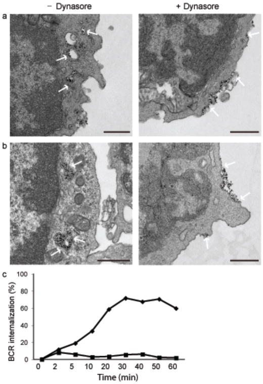
Figure 5. Dynasore blocks BCR internalization.
(a, b) Mouse splenic B cells were treated with 100 μM dynasore in DMSO or DMSO alone for 1 h in serum free RPMI. Cells were incubated with rat antibodies specific for mouse IgM conjugated to magnetic beads for 15 min on ice followed by incubation at 37°C for 10 min (a) or 45 min (b). Cells were fixed and imaged by TEM. Dense black dots represent the BCR indicated by white arrows. Scale bars represent 500 nm. Images are from two independent experiments in which 10–15 grids were examined for each time point.
(c) Mouse splenic B cells were incubated with 100μM dynasore in DMSO (squares) or DMSO alone (diamonds) for 1h in RPMI. Cells were either fixed directly (0 min) or incubated with biotin labeled anti-IgM for indicated times at 37°C. At the end of incubation cells were fixed and stained with streptavidin-FITC and B220-PE. Percent of BCR internalized in B220 positive cells is plotted against time. Data are from one of two independent experiments.

