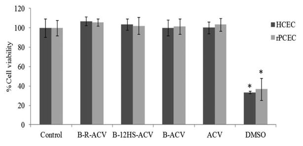
Fig. 6.

Cytotoxicity assay in the presence of B-R-ACV, B-12HS-ACV, B-ACV and ACV on HCEC and rPCEC cells for 48 h. DMSO (10% v/v) served as a positive control. Data represent mean percentage of viable cells ± standard deviation (n= 4). A P-value of less than 0.05 was considered to be statistically significant and denoted by asterisk (*).
