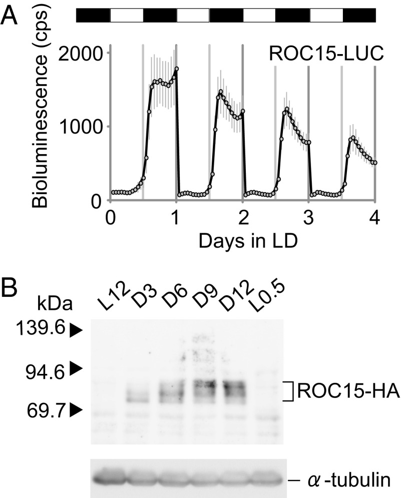
Fig. 1.
Analysis of ROC15 expression in LD. (A) Bioluminescence trace of ROC15-LUC reporter under an LD condition. Asynchronous spot cultures of ROC15-LUC reporter strain was prepared in white 96-well plates as described previously (13). White and black bars represent light (2 μmol⋅m−2⋅s−1) and darkness, respectively. Each point represents the mean ± SD of bioluminescence counts from six independent cultures. (B) Western blot analysis of ROC15-HA protein. Asynchronous high-salt medium cultures (17) of the ROC15-HA strain were exposed to LD conditions, and samples were harvested at the time points indicated. Similar results were obtained in more than five independent experiments and in experiments with another transgenic line (A and B). White light was used in all experiments in this figure.

