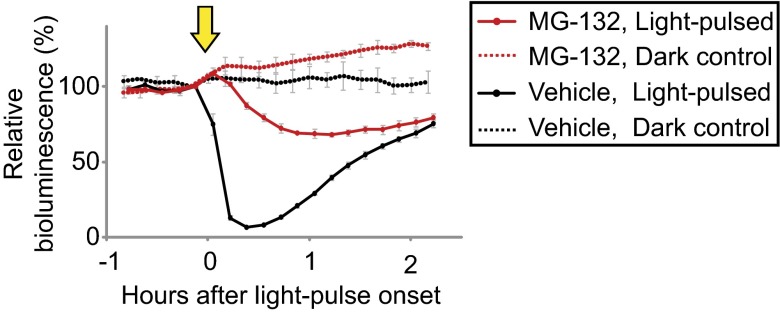
Fig. 3.
The effect of the proteasome inhibitor MG-132. Media were changed to gametolysin-containing media (21, 22) to remove the cell wall for enhanced drug permeability, and then MG-132 was added to a final concentration of 100 μM. After a 30-h treatment in darkness, cells were exposed to a 0.5-min white light pulse (2 µmol⋅m−2⋅s−1, indicated by the arrow). The values just before the light pulses were normalized to 100. Each point represents the mean ± SD of the normalized bioluminescence traces from three independent cultures. Similar results were obtained in three independent experiments and in experiments with another transgenic line.

