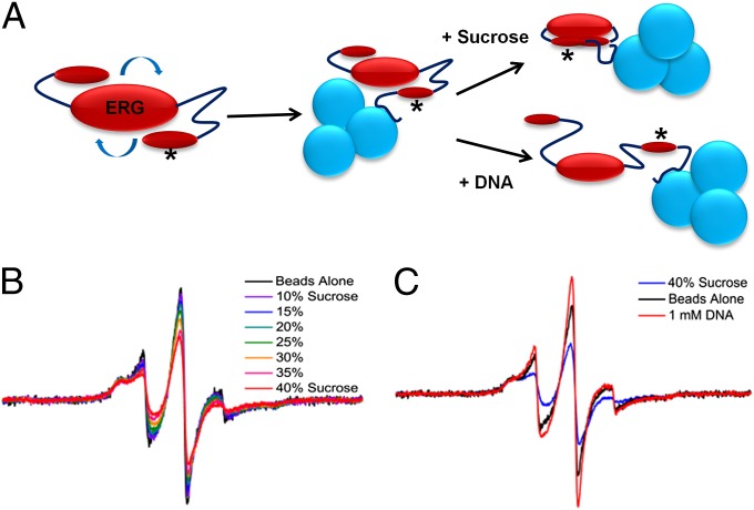
Fig. 2.
EPR experimental setup and results. (A) The rotational correlation time of the fast-tumbling ERG molecule can be greatly reduced by binding the His-tagged protein to Ni-NTA beads before EPR spectroscopy. Asterisk denotes site of MTSL spin label on the ERG NID. (B) Increasing sucrose concentration causes the ERG spin label spectrum to shift from sharp and highly mobile (black trace, no sucrose) to broad and immobile [red trace, 40% (wt/vol) sucrose solution]. (C) Solution containing a high concentration of sucrose shows a shift in the ERG NID toward the ordered state (blue trace), whereas the addition of DNA instead induces the sharper linewidths of the more disordered state (red trace).

