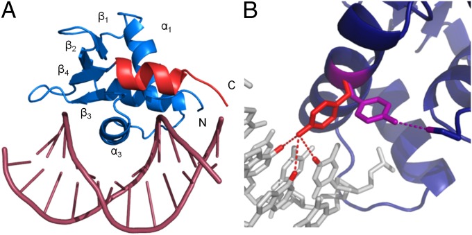
Fig. 4.
Crystal structure of ERGi bound to DNA and change in Tyr354 conformation. (A) ERGi bound to a 12-bp DNA fragment. The Ets domain is shown in blue and the C-terminal autoinhibitory helix in red. No electron density was seen for the N-terminal autoinhibitory region in this structure. (B) Tyr354 on DNA-binding helix a3 in the presence of DNA (red) can form hydrogen bonds with DNA bases Ade7, Ade8, and Thy17. When not bound to DNA (purple), this residue adopts an alternative rotameric state and is positioned to hydrogen bond to Ser283 of the NID, or perhaps other protein binding partners.

