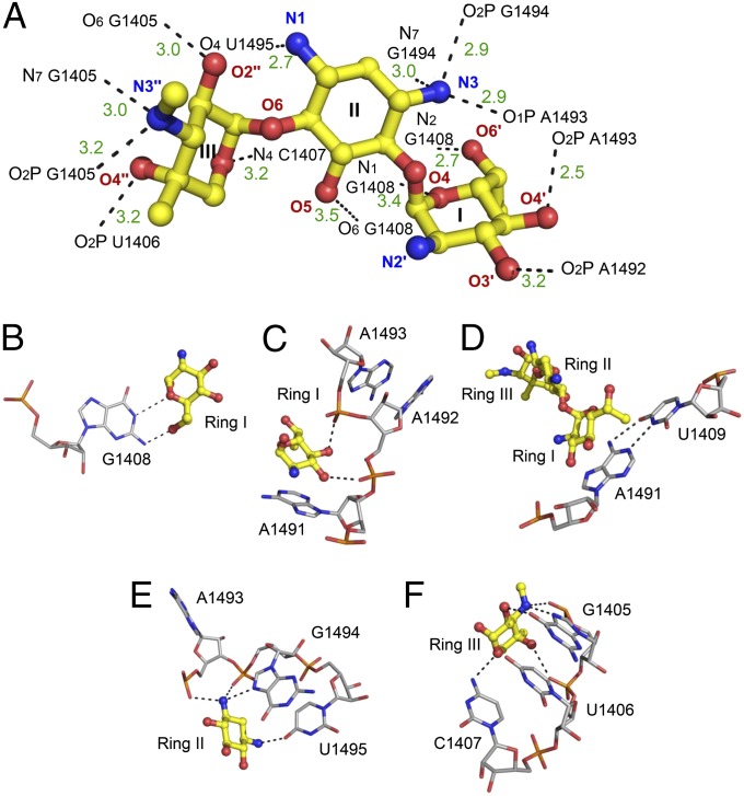
Fig. 4.
Description of the contacts between G418 and the leishmanial A site. (A) The 3D structure of bound G418. Ring numbers (I–III) and atom names are specified. rRNA atoms are numbered according to the E. coli numbering. Hydrogen bonds and salt bridges are presented as black dashed lines. Bond lengths are presented in dark green in ångström (Å). (B–F) The atomic details of the contacts involving the rings with conserved and nonconserved rRNA residues.

