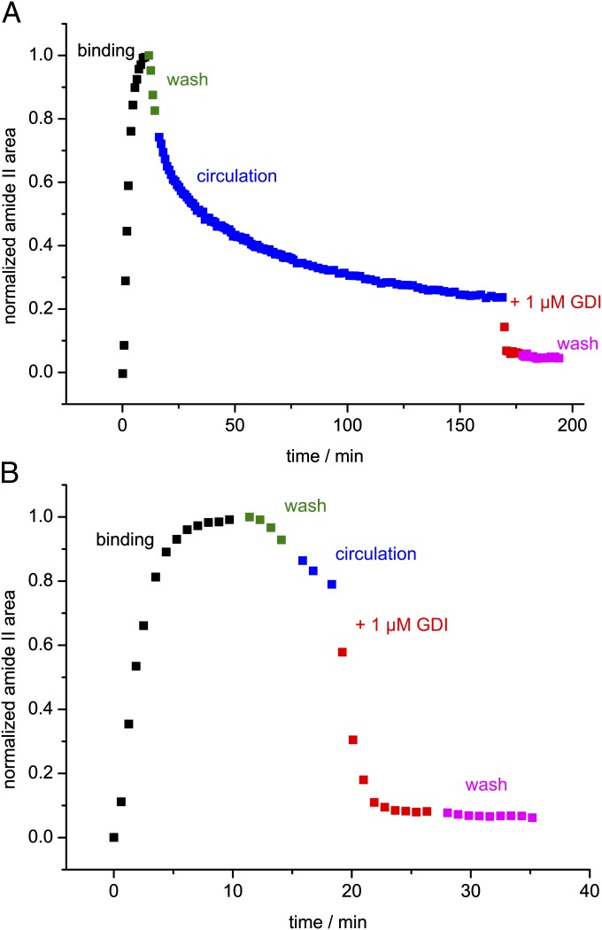
Fig. 3.
Time course of the immobilization of Rab35_G on a lipid bilayer and subsequent extraction by GDI. The experiment consisted of protein binding in a circulating system (black), wash (green), a circulation phase (blue), addition of GDI to the system (red), and a final wash step (magenta). The data points represent the normalized area of the amide II band. (A) An experiment with a prolonged circulation phase. Bound Rab35_G dissociates until equilibrium between the circulating solution and the membrane is reached. Addition of GDI leads to a rapid extraction of the remaining membrane-bound Rab35_G. (B) The same experiment with a short circulation phase. The extraction by GDI is seen more clearly by keeping the circulation phase short.

