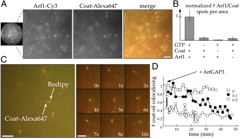
Fig. 2.
COPI produces nanodroplets from LDs. (A) Particles containing Arf1 and coatomer appear in the buffer drops in the presence of the COPI machinery (Left is a full image of a buffer drop; the other three panels are large magnifications to better see the particles). Less than 2 min after making the buffer drops containing Arf1–Cy3 (30 nM), coatomer (15 nM) labeled with Alexa 647, GTP (50 µM), and ARNO (200 nM), homogenous Arf1–Cy3 and coat–Alexa 647 spots appear in the aqueous volume and at the buffer/TAG interface. Arf1 (green) and coat (red) spots are colocalized, moving together in the buffer drop (Movie S1). The spots are slightly separated because of the time delay to switch laser in the setup. (Scale bar, 5 µm.) (B) The formation of particles is GTP-dependent. In the controls without GTP, coatomer or Arf1, the amount of spots per area is significantly reduced compared with the experiment with 50 µM GTP, 30 nM Arf1, and 15 nM coatomer (Left). (C) The particles are TAG nanodroplets. Same experiment as in A with unlabeled Arf1 (100 nM) and Bodipy dye (1% wt/wt) in the TAG. After collection of the buffer drops as indicated in Fig. S1, colocalized Bodipy/Alexa 647 spots are observed. (Scale bars, 10 µm.) (D) Loss of colocalization over time after ArfGAP3 addition. The sample, recovered as shown in Fig. S1, is split in three vials. The amount of particles is quantified as described in Fig. S3. ArfGAP3 is added in two of the samples at different concentrations (50 and 10 nM) corresponding to fractions equal to 0.5 and 0.1 of the Arf1 concentration. Colocalization of coat–Alexa 647 and TAG–Bodipy is lost over time compared with the control.

