Fig. 1.
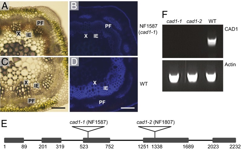
The Medicago cad1-1 mutant shows a lignin deposition defect. (A–D) Cross-sections of stems from sixth internodes of 9-wk-old NF1587 line (cad1-1) and wild-type R108 plants. A and C are light microscopy images. Total lignin in B and D was visualized by UV autofluorescence. IE, interfascicular element; PF, phloem fiber; X, xylem. (Scale bar: 100 µm.) (E) Diagram of the structure of the CAD1 gene and positions of retrotransposon insertions. The numbers indicate nucleotide positions from the site of initiation of translation. The boxes represent exons. The black lines represent introns. (F) RT-PCR analysis of CAD1 transcripts in cad1-1 and cad1-2 mutant and wild-type lines. The actin gene was used as positive control.

