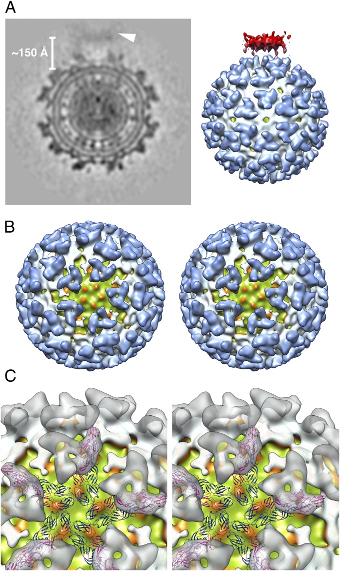
Fig. 5.
Reconstruction of a SINV–liposome complexes when the liposome is associated near the virus icosahedral fivefold axis. (A) Central cross-section (Left) and surface rendering (Right) of the reconstruction map. The target membrane (arrowhead) is ∼150 Å away from the outer leaflet of the viral membrane. The surface-shaded view of the reconstruction map is contoured at the 1σ level. The coloring scheme for the virus components is consistent with Fig. 1A, with the target membrane colored in red. (B) Liposome-binding site on the virus (toward the reader) along the fivefold axis. The reconstruction map was rendered at the 1.5σ contour level. Five bulging densities on the outer membrane leaflet of the virus (green) are colored orange. (C) Enlarged view of the reconstruction map with fitted CHIKV virus E1 (blue) and E2 (magenta) glycoproteins (17). For clarity, the virus spikes are represented as transparent gray surfaces.

