
Fig. 1.
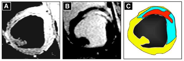
Visualization of cardioprotection by magnetic resonance imaging (MRI). MRI short axis images obtained at the same left ventricular level 3 days after MI induction in a pre-reperfusion-metoprolol-treated animal. Panel A shows a T2-weighted, fast spin-echo image, where the hyperintense area indicates the presence of edema. Panel B shows a delayed enhancement image after contrast administration, depicting the infarcted area (bright). In panel C, the area at risk (edema, blue) and infarcted area (red inside the blue region) shown in panels A and B are merged. Non-ischemic myocardium is shown in yellow. Note the large salvaged myocardium (blue area surrounding the infarcted red zone).