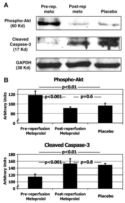
Fig. 4.
Protein expression of markers of reperfusion injury. Western blotting (Panel A), and its quantification by densitometric units (Panel B, expressed as mean±standard error of the mean) of phospho-Akt and cleaved caspase-3 in animals allocated to the pre-reperfusion-metoprolol, post-reperfusion-metoprolol and placebo. See also text. Glyceraldehyde-3-phosphate dehydrogenase (GAPDH). N=12 (4 per group).

