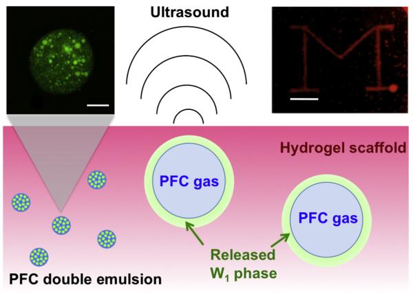
Fig. 1.
Schematic representation of drug release by ADV in a hydrogel scaffold. Bottom: A water-in-PFC-in-water (W1/PFC/W2) double emulsion, containing a growth factor in the W1 phase, is encapsulated within the scaffold. Upon exposure to acoustic amplitudes greater than the ADV threshold of the emulsion, the PFC within the droplets is vaporized, thus releasing the W1 phase. Top left: confocal fluorescence image (fluorescein, shown in green) of a PFC double emulsion droplet (scale bar = 10 μm). Smaller aqueous droplets, containing a water-soluble payload such as fluorescein or bFGF, are enveloped by a larger PFC globule. Top right: visible image of a 10 mg ml−1 fibrin gel containing 5% (v/v) double emulsion after targeted exposure to ultrasound. The “block M” consists of gas bubbles generated by ADV. Scale bar = 4 mm.

