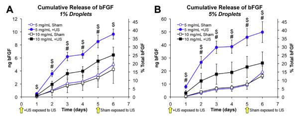
Fig. 5.

ADV-induced release of bFGF from droplet–hydrogel composites.+US groups were exposed to ultrasound at the start of the experiment (i.e. time = 0 days) and shams were exposed to ultrasound after collection of releasate on day 5. Cumulative release profiles are shown in (A) and (B). Blue series indicate 5 mg ml−1 fibrin and black series indicate 10 mg ml−1 fibrin. Data are shown as mean ± standard deviation for n = 5. $p < 0.05 for 5 mg ml−1 fibrin + US vs. sham control. #p < 0.05 for 10 mg ml−1 fibrin + US vs. sham control. For sham controls with 5% emulsion (for both 5 mg ml−1 and 10 mg ml−1 fibrin) treated with ultrasound at day 5, rates of release were found to be significantly higher at day 6 compared to projected values computed using the 95% confidence interval of the slope between days 1–5.
