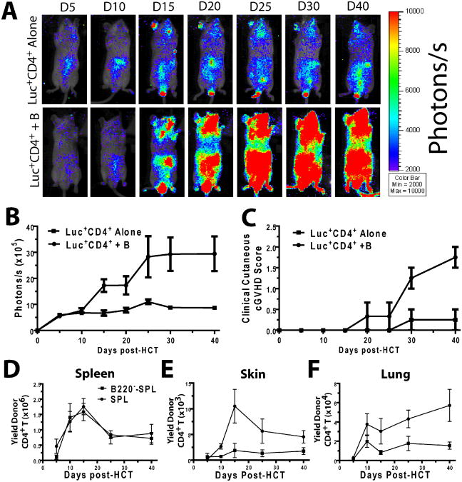
Figure 6. Donor B cells in transplants augmented donor-type CD4+T cell expansion in GVHD target tissues.
A-C. BALB/c mice were irradiated (800 cGy) and injected with 5×10⁶ CD4+CD25− splenoctyes from luciferase transgenic (luc+) DBA/2 mice and WT DBA/2 TBCD SPL (5×10⁶), with or without WT DBA/2 B220+ B cells (10×106), then monitored for expansion of donor CD4+ T cells and signs of GVHD. A. CD4+ T cell expansion in mice was evaluated with bioluminescent imaging (BLI). One representative BLI pattern is shown per group per time point (n=8 from two replicate experiments). B. BLI intensity in terms of photons/s. A summary curve (mean±SE) is shown. Mice recieiving B cells had increased luminescence (2-way ANOVA, p<0.01). C. Recipieints of luc+CD4+ T cells with or without B cells were evaluated after HCT for GVHD-related skin damage and hair loss. Recipients of B cells had increased clinical cutaneous scores (2-way ANOVA, p<0.01). D-F. BALB/c mice were irradiated (800 cGy) and injected with either SPL or B220−-SPL and sacrificed 5, 10, 15, 25, and 40 days post-HCT, and their tissues were harvested and evaluated for the presence of infiltrating donor CD5.1+CD4+ T cells. Mean±SE is shown at each time point, n=4-8 from 2-3 replicate experiments. D. Donor CD4+ T cell yield in the spleen was evaluated, and was not significantly different between SPL and B220−-SPL groups (2-way ANOVA, p>0.1) E. Donor CD4+ T cell yield in the skin was evaluated. A higher yield was observed in SPL recipients (2-way ANOVA, p<0.01). F. Donor CD4+ T cell yield in the lung was evaluated. A higher yield was observed in SPL recipients (2-way ANOVA, p<0.01).

