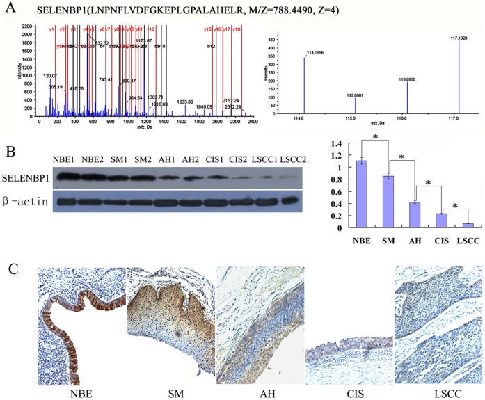
Figure 1. Expressional changes of SELENBP1 in the human bronchial epithelial carcinogenic process.
A, MS/MS spectra used for the identification and quantitation of SELENBP1. (left) the sequence LNPNFLVDFGKEPLGPALAHELR allows the identification of SELENBP1. (right) the released iTRAQ reporter ions provide the relative quantitation of SELENBP1 from the four tissues evaluated. NBE, labeled with iTRAQ reagent 117; SM, labeled with iTRAQ reagents 114, AH/CIS, labeled with iTRAQ reagent 116; and invasive LSCC, labeled with iTRAQ reagent 115. B, detection of SELENBP1 expression in the microdissected tissues by Western blotting. (left) a representative result of Western blotting shows the expressions of SELENBP1 in the microdissected NBE, SM, AH, CIS and invasive LSCC; (right) histogram shows the expression levels of SELENBP1 in these tissues as determined by densitometric analysis. β-actin is used as the internal loading control. Columns, mean from 10 cases of tissues; bars, S.D. (*, P<0.05 by One-way ANOVA). C, detection of SELENBP1 expression in the formalin-fixed and paraffin-embedded archival tissue specimens by immunohistochemistry. A representative result of immunohistochemistry shows the expression of SELENBP1 in the NBE, SM, AH, CIS and invasive LSCC. Original magnification, ×200.

