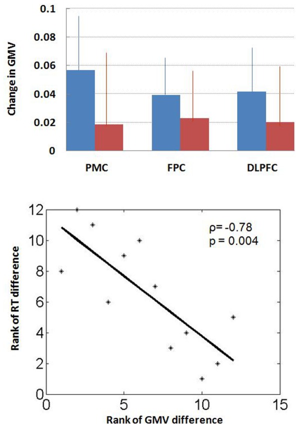
Figure 2. Changes (mean ± s.d.) in Gray matter volume (GMV) for the three regions of interest: primary motor cortex (PMC), frontopolar cortex (FPC), and dorsolateral prefrontal cortex (DLPFC), for the ADT (blue) and control (red) group (upper panel).

A decrease in the GMV of the PMC correlated with prolonged reaction time to target detection during the zero-back condition in the ADT group (lower panel). Because of the small sample size, we used a Spearman regression for correlation (p<0.0042, rho = −0.7832). The result of Pearson regression was also significant: p<0.0049.
