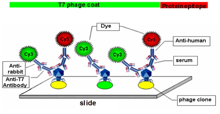
Figure 1. Dual-color fluorescent protein microarray detecting system.
Sera from MM patients and normal donors were used as the source of primary antibodies for detecting phage-expressed, immunogenic proteins. Mouse anti-T7 antibody was used to detect the T7 phage capsid proteins as an internal control. Fluorescently labeled Cy5 anti-human and Cy3 anti-mouse secondary antibodies were used to visualize the primary antibodies. Each spot on the slide had a green signal from the T7 phage capsids, while the immunogenic phage clones had red signals from serum autoantibodies.

